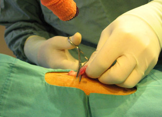
| The laser is shown making the initial skin incision. |
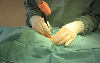 |
| Note how small the incision is. |
 |
| The laser is also used to go through the fatty tissue under the skin, and finally through the muscle layer leading into the abdomen. |
 |
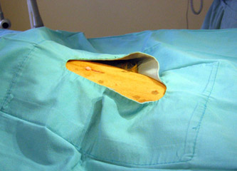
| A special instrument is then used to locate the uterus and bring it through the incision. |
 |
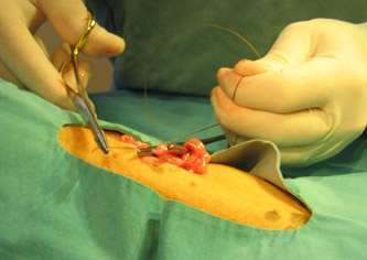
| The first ovary is exteriorized.ĀThe blood supply to this ovary is then clamped off. |
 |
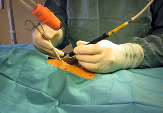
| A second clamp is applied, then the blood supply to the ovary is ligated or tied using a special dissolving suture material. |
 |
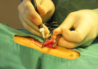
| The laser then vaporizes the blood vessels leading to the ovary, allowing the ovary to be separated and the tied off blood vessels to be released safely back into the abdomen. |
 |
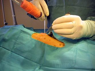
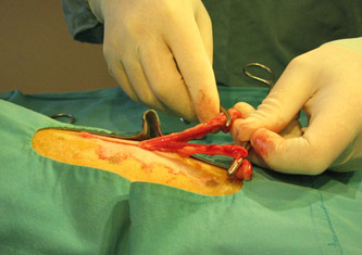
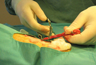
| The horn of the uterus is then followed down to the body, then up to the horn and the ovary on the opposite side.ĀThis ovary and horn are then separated from the blood supply in a similar fashion to the first ovary and horn.ĀIn this picture, the Y-shaped uterus is shown, with the ovaries just beyond the two surgical clamps.ĀThis entire structure will be removed. |
 |
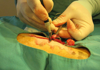
| The body of the uterus along with the uterine blood vessels are clamped, then tied with an absorbable suture material. |
 |
| Next the uterus is removed using the surgical laser. |
 |